
A full set of adult teeth has 32 teeth. Many of today's mouths have comfortable room for 28. The four extra are always the same ones: the third molars, the wisdom teeth, which arrive last (between ages 17 and 25) and find the row already full.
From there, anything can happen. Some come in straight and work for a lifetime. Others come halfway through and get inflamed every so often. Others stay lying inside the bone without giving a single sign for years. And around all of them circulates the same dinner-table advice: "get them out before they make your teeth crooked".
That advice, precisely, is the one the evidence doesn't support. In this article we explain how the decision is really made: when it makes sense to take them out, when it's better to leave them and monitor them, what it means for a tooth to be impacted, what the extraction is like in each case and what to expect in the following days.
In short
Not every wisdom tooth is taken out. They're extracted when they cause problems or are going to: repeated gum infections, decay in them or in the molar in front, cysts, damage to the neighboring root, or when an orthodontic plan requires it. An impacted wisdom tooth is one that couldn't come in (it stayed under the gum or inside the bone), and a partially impacted one is one that only came through in part. The most recent Cochrane review concludes that there isn't enough evidence to routinely remove those that are healthy and symptom-free, and that taking them out doesn't prevent crowding. What does matter is timing: surgery after age 25 is associated with more complications. And if they're left, they're monitored.
Why do wisdom teeth cause so many problems?
Because of space and the order they arrive in. They're the last teeth to form and come in, and they do so at the back of the arch, where the bone curves upward and ends. If there isn't enough room, the tooth does what it can: it comes in tilted, comes in halfway or doesn't come in at all.
The problem isn't the tooth itself, but the position it ends up in. A straight, fully erupted wisdom tooth is brushed like any other. A tilted tooth, or one half-covered by gum, creates a corner no toothbrush can reach, right against the molar in front, which you really do need.
What does it mean for a wisdom tooth to be impacted?
It's the question that causes the most confusion, because the terms are used loosely. This is the classification we use at the clinic:
- Erupted. It came in completely, sits in the arch at the same height as the others, and bites against the tooth above or below. For all intents and purposes, it's just another molar.
- Partially impacted. Only part of the crown came through; the rest is still covered by gum or bone. It's the situation that causes the most infections, because it leaves a door ajar between the mouth and the space around the tooth.
- Impacted. It didn't come in, even though the age when it should have has passed. It may be covered only by gum (soft tissue impaction) or enclosed in bone (bony impaction). And it may be vertical, tilted forward, tilted backward or horizontal, which is when it lies against the root of the neighboring molar.
A point worth clarifying: a wisdom tooth you can't see isn't necessarily impacted. At 15, a wisdom tooth that hasn't come in simply hasn't finished forming yet. It's called impacted when the root has formed, the eruption age has passed, and the X-ray shows it has no path to come in. That's why the diagnosis isn't made by looking in the mouth, but with a panoramic X-ray.
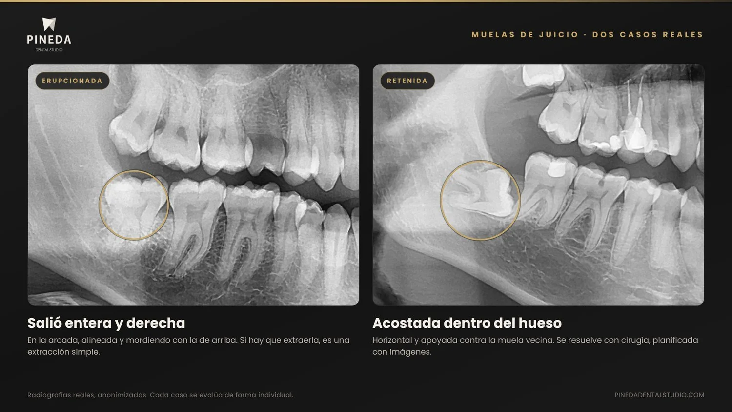
Wisdom teeth · Pineda Dental Studio
Three situations, three approaches
The same tooth may need nothing, monitoring or surgery. What decides which is the position, not the fact of having it.
| Situation | What it looks like | Typical risk | If it has to come out | Usual approach |
|---|---|---|---|---|
| Erupted Came in completely and aligned. | Fully visible in the mouth, at the same height as the others. | LowIf it can be cleaned. | Simple extraction, similar to any other molar. | Conservarla y controlarla. Se extrae si tiene caries no restaurable o no se logra limpiar. |
| Partially impactedThe most troublesome Only came through in part. | One tip of the crown is visible; the rest is under a flap of gum. | HighGum infection and decay in the neighboring molar. | Surgical extraction of low to medium complexity. | It's the one most often extracted, especially after a second episode of infection. |
| Impacted Didn't come in: under the gum or inside the bone. | Not visible in the mouth. It only shows up on the X-ray. | VariableDepends on its position and what's nearby. | Surgery, with imaging of its relationship to the nerve. | Remove it if there's disease or a risk to the neighboring molar. If it's deep, healthy and symptom-free, it can be monitored. |
"Impacted" doesn't mean "it needs surgery". It's a description of where the tooth is. The indication comes from what that position is doing, or could do, to what's around it.
Classification and approach are decided with a panoramic X-ray and, when needed, a CT scan.
How do I know if I need them out?
There are signs you can notice yourself and findings that only show up on the X-ray. Both lists matter.
What you may notice
- Pain and swelling of the gum at the back of the mouth, which comes, eases and returns. It's called pericoronitis: infection of the flap of gum covering a half-erupted tooth. Sometimes it comes with a bad taste, difficulty opening the mouth or swollen glands.
- Food that always gets stuck in the same place, behind the last molar.
- Repeatedly biting your cheek, when the upper tooth came in tilted outward.
- Pain that spreads to the ear or temple on the same side.
What the X-ray shows
- Decay in the wisdom tooth that can't be restored properly because of its position.
- Decay on the back of the neighboring molar, right where the wisdom tooth rests.
- Bone loss between the two molars.
- Root resorption of the neighboring molar from the pressure.
- Un quiste alrededor de la corona de la muela retenida.
On top of that, there are indications from a treatment plan: when orthodontics, a prosthesis or surgery needs that space, or when the tooth has nothing to bite against and starts to over-erupt.
If you recognize yourself in the first list, it needs looking at. If you have no symptoms, a panoramic X-ray around age 17 or 18 is still a good idea: it's the time when you can see where they're heading, with time to decide.
Does taking them out keep my teeth from getting crooked?
No. It's the most repeated reason for removing them and the one with the least support.
The idea is intuitive: the wisdom teeth push from behind and crowd the front teeth. But when it was put to the test, it didn't hold up. The Cochrane review on the subject (the most rigorous reference there is) included a clinical trial that measured teenagers' arches for five years and found no clinically significant effect of extraction on crowding of the incisors (Ghaeminia et al., 2020).
Front teeth tend to crowd over the years in almost everyone, with or without wisdom teeth; it's part of how the mouth changes with age, and that's why retainers are used after orthodontics. We cover it in the article on types of orthodontics. If someone tells you to take out all four wisdom teeth just "so your teeth don't move", it's worth asking for a second reason.
What if I have no symptoms? Should I take them out just in case?
This is where thinking has changed most in recent years, and where we prefer to be clear about what's known and what isn't.
The same Cochrane review looked at all the available evidence on impacted, healthy, symptom-free wisdom teeth and concluded that there isn't enough evidence to determine whether it's better to remove them or keep them. It found, with very low certainty, that keeping them might increase the long-term risk of gum disease in the neighboring molar; and it recommended deciding case by case, with regular check-ups when they're left in (Ghaeminia et al., 2020).
In practice, that rules out two extremes. Removing all four wisdom teeth from everyone at 18 has no support. Leaving them and forgetting about them doesn't either. What we do is assess the position: an impacted tooth that's deep, fully covered by bone and away from its neighbor can stay, with an X-ray every so often. A tooth tilted forward and touching its neighbor is another story, even if it doesn't hurt today.
What can happen if I leave them?
It depends almost entirely on the position. These are the problems we see, from most to least common:
- Recurrent pericoronitis. Episodes tend to repeat, and an infection that progresses can spread to nearby tissues. A first mild episode is treated; when it repeats, that's usually the indication to extract.
- Decay in the neighboring molar. It's the damage that concerns us most, because it affects a molar you do need. A meta-analysis found decay on the back of the neighboring molar in about 36% of wisdom teeth tilted forward and 22% of horizontal ones (Tödtling, 2023). It's decay that's hard to see and hard to restore.
- Bone loss and a periodontal pocket behind the neighboring molar. We treat it in our gum treatment area, but it's better not to get there.
- Resorption of the neighboring root, from sustained pressure.
- Cysts around the crown of an impacted tooth. Uncommon, and silent: they show up on the follow-up X-ray.
And nothing may happen at all. That's why, when the decision is to keep them, the key word is monitoring.
Does age matter?
Yes, and it's the strongest argument for not postponing an extraction that's already indicated.
Between ages 17 and 25, the wisdom tooth's roots haven't finished forming, the bone is more elastic and recovery is faster. Over the years the roots finish forming and curve, the bone becomes denser and the tooth ends up closer to the nerve. A study of more than 4,000 patients and 8,700 third molars found that patients over 25 were about 46% more likely to have complications than younger ones (Chuang et al., 2007).
That doesn't mean everyone should have surgery at 18. It means that if the X-ray shows a tooth that's clearly going to cause problems, waiting until it hurts at 35 isn't the best strategy.
What about the nerve?
It's the most common fear among people about to have their lower wisdom teeth out, and it deserves an answer with numbers.
Inside the lower jaw runs the inferior alveolar nerve, which gives feeling to the lip and chin on that side. The roots of a lower wisdom tooth can be very close to it. If the nerve is irritated during surgery, a numb or tingling feeling is left in the lip.
A review of the literature on the subject found an incidence of between 0.35% and 8.4%, and that 96% of cases recover within four to eight weeks; permanent changes are uncommon. It also identified the factors that increase the risk: age over 24, teeth in a horizontal position and radiographic closeness between the root and the nerve canal (Sarikov & Juodzbalys, 2014).
That's why, when the panoramic X-ray shows the root is close to or overlapping the canal, we ask for a CT scan before surgery. The panoramic is a flat image; the CT scan shows in three dimensions whether the nerve runs in front of, behind or between the roots, and that's used to plan how to approach the tooth. And there are cases where, after seeing the CT scan, the best decision is to leave it alone.
Wisdom teeth · What the evidence says
Three numbers to help you decide
Extraction isn't as risky as people fear, and waiting isn't always free.
Of changes in sensation from the inferior alveolar nerve after extraction (Sarikov & Juodzbalys, 2014).
In a study of more than 4,000 surgical patients (Chuang et al., 2007).
Or more, versus 1–5% for a routine extraction. With chlorhexidine the odds drop by more than half (Daly et al., 2022).
All three can be worked on: the first, with a CT scan and planning; the second, by not postponing what's already indicated; the third, with care during the first week.
What is the extraction like?
It depends on which of the three situations is yours, and that's why the visit beforehand matters so much.
An erupted wisdom tooth is extracted like any other: local anesthesia, it's loosened and removed. It usually takes a few minutes and the recovery is mild.
A partially impacted or impacted wisdom tooth is surgery, which at Pineda Dental Studio is performed by our maxillofacial surgery team. Under local anesthesia, a small window is opened in the gum, the bone covering the tooth is removed, and the tooth is often divided into two or three parts to take it out through a smaller space and touch less bone. It's closed with stitches. With the area numb, what you feel during surgery is pressure and vibration.
As for how many to take out at once: when it makes sense, we remove all four in one session, which concentrates the recovery into a single week. In other cases we prefer to do one side at a time, so there's always a side to chew with. We decide based on how complex each tooth is and what will be easier for you.
At the end we apply low-power laser (photobiomodulation) to the area, with the aim of reducing inflammation and discomfort over the following days. It's the same unit we use in other treatments, which we describe on our laser dentistry page.
What are the following days like?
Recovery follows a fairly predictable course. Knowing it in advance avoids half the scares.
- The first few hoursBite on the gauze with firm pressure for 30 to 45 minutes. Cold on the outside, at intervals. No rinsing, spitting or straws: all of that dislodges the clot that needs to stay in place. Cold or lukewarm, soft food. No smoking.
- Days 1 and 2Expect moderate discomfort, which eases with the prescribed medication, and swelling that increases. Sleeping with your head slightly raised helps. Brush the rest of your mouth as usual.
- Day 3This is when swelling peaks, and a bruise may appear on the cheek. It's alarming, but it's normal: from here it should start going down. Cold is usually switched for warmth.
- Days 4 to 6Swelling goes down and your mouth opens wider each day. If instead the pain starts rising again on these days, it may be a dry socket: we explain below how to recognize it.
- Day 7Check-up and removal of stitches, if they're not dissolvable. Most people are back to their routine.
- The following weeksThe gum closes in two to three weeks. The bone takes months to fill the space, but you don't feel that. Meanwhile, rinse the area gently after eating.
What dry socket is and how to prevent it
It's the most common complication, and the most uncomfortable. It happens when the clot that protects the bone is lost or breaks down, leaving the bone exposed. You can recognize it because the pain, instead of going down, increases on the third or fourth day, spreads toward the ear and doesn't ease with the usual painkillers; sometimes there's a bad taste or smell.
A Cochrane review of 49 clinical trials notes that it occurs in 1% to 5% of routine extractions and in 30% or more of surgically removed third molars, and found moderate-certainty evidence that chlorhexidine prevents it: gel placed in the socket reduced the odds of dry socket by 58% (Daly et al., 2022). The other factor that weighs most depends on you: not smoking in the following days, and protecting the clot in the first few hours.
If it happens, it's not serious, but it does need treatment: the socket is cleaned and a local medication is placed that brings immediate relief. Don't wait for it to go away on its own.
When to message us right away
- Bleeding that doesn't stop after biting on gauze with pressure for 30 minutes.
- Pain that increases after the third day instead of decreasing.
- Swelling that keeps growing after the fourth day, or fever.
- Numbness of the lip or chin that lasts beyond the effect of the anesthesia.
- Difficulty swallowing or breathing: that doesn't wait for an appointment, it's an emergency room visit.
For blows, broken teeth or teeth knocked out of place, you'll find the full guide in what to do in a dental emergency.
Frequently asked questions
At what age do wisdom teeth come in?
Generally between ages 17 and 25. Some people get them later, some never get them, and some simply never formed one or more.
Do all wisdom teeth have to come out?
No. Those that came in completely, are aligned, can be cleaned and have no decay are kept like any other molar.
What's the difference between impacted and partially impacted?
An impacted tooth didn't come in: it's under the gum or inside the bone and only shows on the X-ray. A partially impacted one came through in part. The second is the one that gets infected most often.
Will I be put to sleep for the surgery?
At the clinic it's done under local anesthesia, the same as for any dental treatment. With the area numb, what you feel is pressure and vibration.
How many days of rest do I need?
After a simple extraction, one or two. After surgery on impacted wisdom teeth, it's best to set aside three to five days, knowing that swelling peaks on the third.
Should I have all four out at once?
It depends on the complexity. Doing it in one session concentrates recovery into one week; doing one side at a time always leaves a side to chew with. We decide at the visit.
Could I lose feeling in my lip?
It's a known risk with lower wisdom teeth close to the nerve, with a low incidence and recovery within a few weeks in the vast majority of cases. That's why a CT scan is requested when the X-ray shows the tooth is close.
I have an impacted wisdom tooth that has never bothered me. Should I leave it?
That may be the right decision, if it's deep, healthy and away from the neighboring molar. The condition is monitoring it with regular X-rays. What isn't advisable is leaving it unchecked.
The decision is made with an X-ray, not with advice
Wisdom teeth aren't a problem just for existing. They become one when their position puts something else at risk: the gum, the bone or the molar next to them. Knowing which is your case takes a panoramic X-ray and a conversation. If you're looking for a dentist in Recoleta or anywhere in Buenos Aires to assess your wisdom teeth, at our dental clinic we'll tell you which ones should come out, which can stay, and why.
Ayacucho 1230, Recoleta · We reply the same day
📍 We look forward to seeing you at our clinic in Recoleta, Buenos Aires.
👩🏻⚕️ About the author and clinical director
Od. Mariana Pineda (M.N. 39.080) is a dentist (José Antonio Páez University, 2014) and trained in fixed, removable and implant-supported prosthodontics at the Argentine Dental Association (2018). As clinical director of Pineda Dental Studio, she leads a multidisciplinary team of more than 10 dentists in maxillofacial surgery, implants, oral rehabilitation, cosmetic dentistry and endodontics, following international protocols for minimally invasive dentistry.
Medical notice: the information in this article is for educational purposes. Whether a wisdom tooth should be removed or kept, the technique and post-operative care depend on the individual clinical and X-ray diagnosis we make at the clinic.
📚 Scientific sources consulted
- Ghaeminia, H., Nienhuijs, M. E. L., Toedtling, V., Perry, J., Tummers, M., Hoppenreijs, T. J. M., Van der Sanden, W. J. M., & Mettes, T. G. (2020). Surgical removal versus retention for the management of asymptomatic disease-free impacted wisdom teeth. Cochrane Database of Systematic Reviews, 2020(5), CD003879.
- Daly, B. J. M., Sharif, M. O., Jones, K., Worthington, H. V., & Beattie, A. (2022). Local interventions for the management of alveolar osteitis (dry socket). Cochrane Database of Systematic Reviews, 2022(9), CD006968.
- Sarikov, R., & Juodzbalys, G. (2014). Inferior alveolar nerve injury after mandibular third molar extraction: a literature review. Journal of Oral & Maxillofacial Research, 5(4), e1.
- Chuang, S. K., Perrott, D. H., Susarla, S. M., & Dodson, T. B. (2007). Age as a risk factor for third molar surgery complications. Journal of Oral and Maxillofacial Surgery, 65(9), 1685–1692.
- Tödtling, V. (2023). Second molar distal surface caries in the context of mandibular third molar absence and impactions [tesis doctoral]. Vrije Universiteit Amsterdam. doi:10.5463/thesis.130
